


?What is EBUS? Why use this method
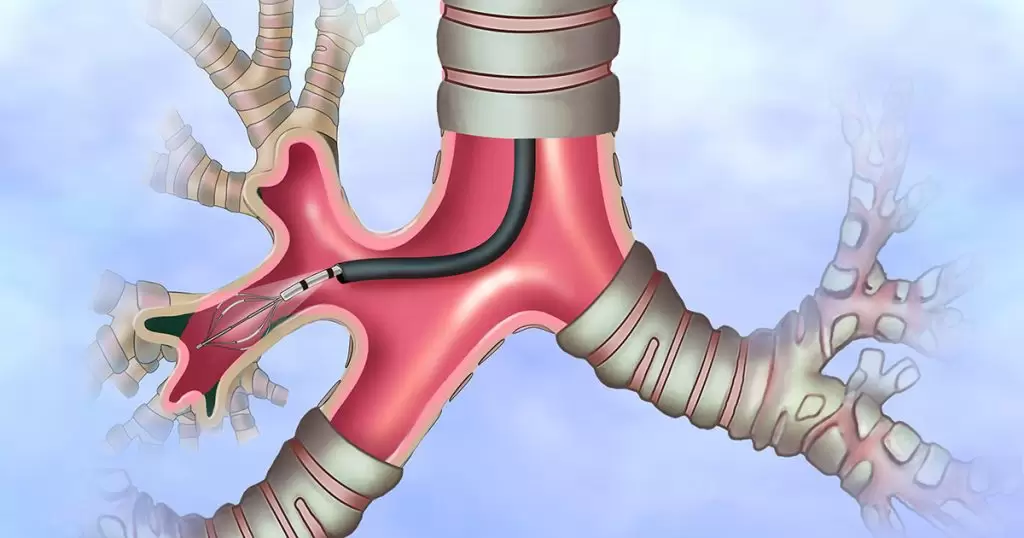
EBUS bronchoscopy (endobronchial ultrasound) is used to diagnose different lung disorders such as infection, cancer, or inflammation. EBUS bronchoscopy is performed by a pulmonologist using a flexible tube that passes through your mouth and enters the trachea and lungs. Dr. Arda Kiani performs this procedure in the best hospitals in Tehran, Iran.
What is the application of EBUS?
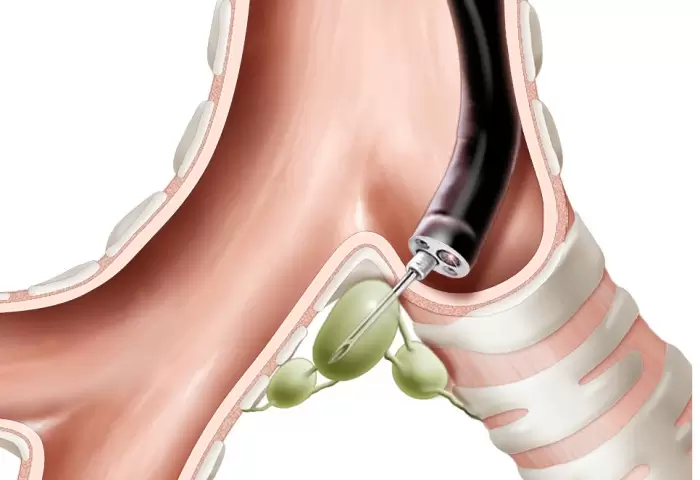
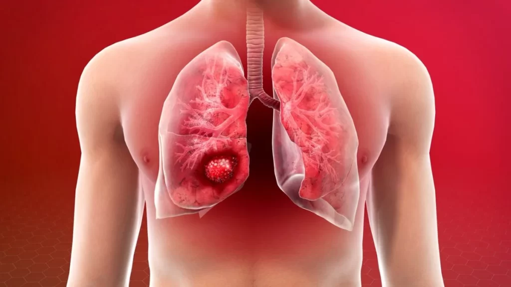
EBUS allows doctors to perform a technique called transbronchial needle aspiration (TBNA) to get fluid or tissue samples from lymph nodes or lungs without surgery. The samples can be used to diagnose and determine the stage of pulmonary cancer, diagnose infections, and detect inflammatory diseases affecting the lungs including sarcoidosis and cancers like lymphoma.
The main indications for endobronchial ultrasound in clinical practice include:
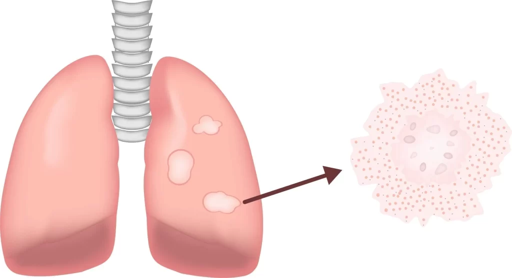
- Diagnosing central lung parenchymal lesions
- Diagnosing mediastinal lesions
- Neoformation or Lymphadenopathy of an unspecified nature
- At the Mediastinal stage before lung cancer surgery
- Lung cancer Mediastinal reconstruction after neoadjuvant radiotherapy/chemotherapy
EBUS thus provides the chance for an accurate diagnosis of cancer and detection of its stage through a systematic mediastinal lymph node study.
This technique is used in other pathological diagnoses including:
- Lymph node or other organs’ metastasis
- Sarcoidosis
- Tuberculosis
- Lymphomas
- Other infections
What sets EBUS apart?
The conventional diagnostic procedure entails a surgery called mediastinoscopy to access the chest. A small incision is made in the neck next to or above the breastbone. Then, a thin scope (mediastinoscope) is inserted through the incision to access the lungs and surrounding lymph nodes. Fluid or tissue is then collected through a biopsy.
What are the advantages of EBUS?
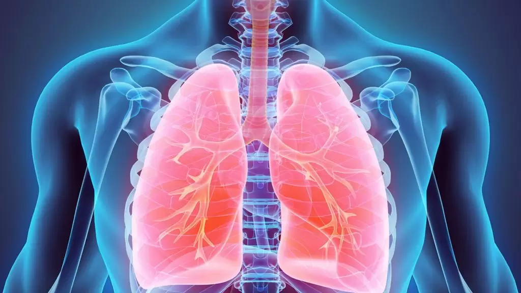
- Real-time imaging of blood vessels, lungs, airways, and lymph nodes
- Improved images allow the doctor to better view hard-to-see sites and access smaller and more lymph nodes for needle aspiration biopsy compared to conventional mediastinoscopy
- The speed and accuracy of EBUS allow quick pathological evaluation on site
- Is performed under moderate sedation or general anesthesia
- Patients recover quickly and can typically be discharged the same day
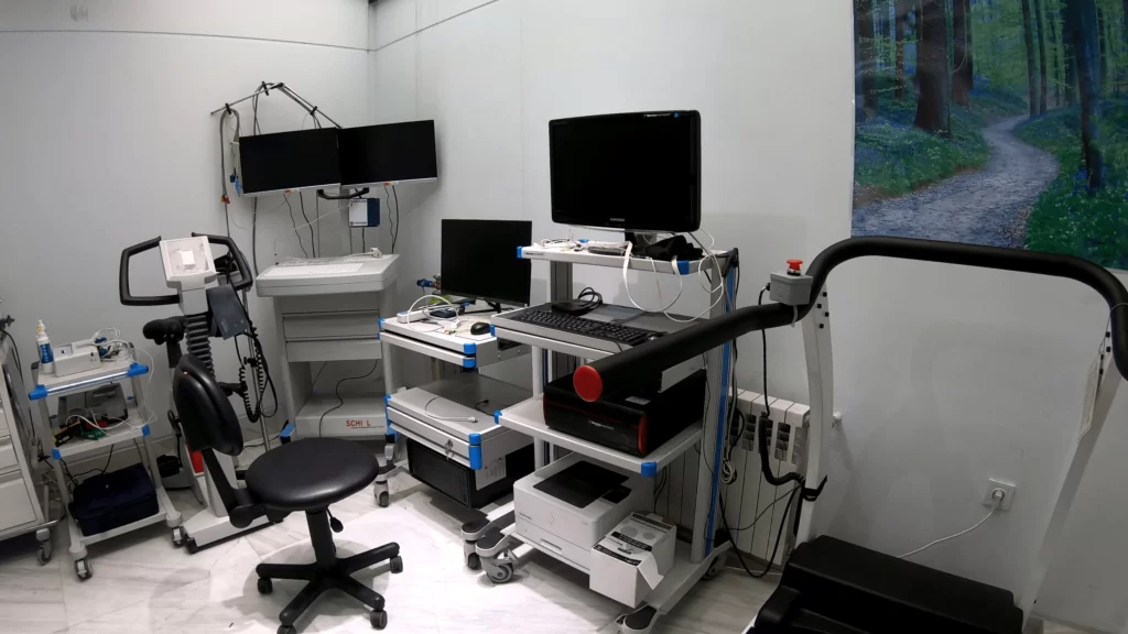
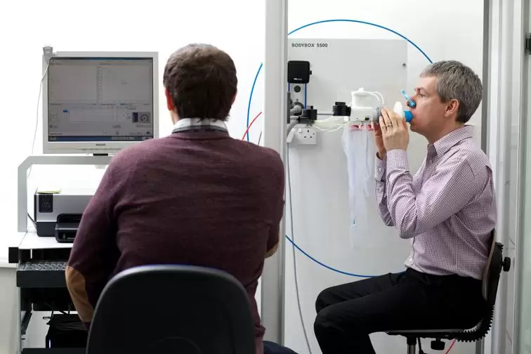
EBUS is performed in hospitals under deep sedation (similar to general anesthesia) and takes between 20 minutes and one hour depending on the doctor’s opinion. This examination requires preparatory tests (respiratory function tests, electrocardiogram, and blood tests,) and referral to a pulmonologist and anesthesiologist. The patient must fast for at least six hours before the surgery. It would be best to have a companion to go back home since the procedure is performed under deep anesthesia.
-

problems of patients and the medical community
27 خرداد 1402 -
Lung clinic
8 خرداد 1402 -

Long COVID
8 خرداد 1402 -
Sarcoidosis Clinic
8 خرداد 1402 -
?What is lung cancer, and how is it treated
8 خرداد 1402 -
Sarcoidosis and pulmonary fibrosis
8 خرداد 1402 -
What exactly is foreign body aspiration?
8 خرداد 1402 -
How are pulmonary function tests؟
8 خرداد 1402 -
Everything you require to know about the polysomnography
8 خرداد 1402 -

?How is pulmonary rehabilitation carried out
8 خرداد 1402